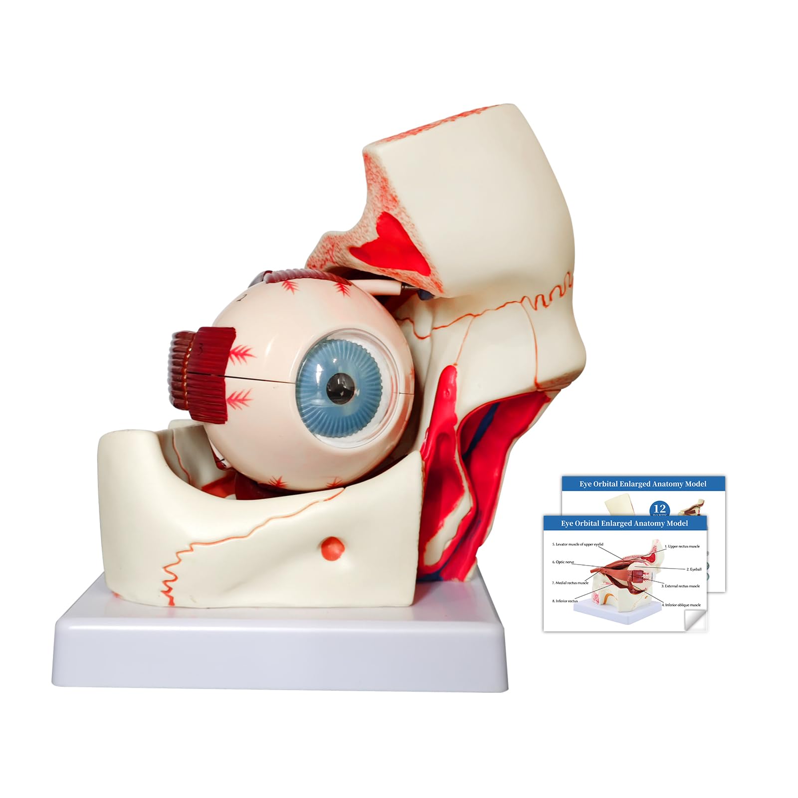
Anatomically Accurate - This is a 12-part, triple-magnified anatomical model of the eyeball orbital anatomy, including the following removable parts: Orbits, sclera of the eyeball wall, superior and inferior hemispheres, lens, vitreous humor, and extraocular muscles and optic nerves Widely used - The model finds utility in science education, student learning, display purposes, and medical teaching. It caters to professionals such as physiotherapists, radiology technicians, and medical practitioners. Its adaptability makes it suitable for various educational and medical environments High-Quality Construction - Constructed from non-toxic PVC, high-strength, realistic in shape, light and strong and easy to disassemble and assemble. The model is environmentally friendly, corrosion-resistant and long-lasting. Its realistic design is both lightweight and sturdy, ensuring ease of handling and assembly Professional Educational Tool - This eye model serves as an effective educational tool suitable for medical training, science classes, and professional development. It accurately represents the anatomical structure of the human eye, emphasizing key features such as the three layers of the eye wall and major refractive components Portable 3D Mannequin - The orbital eye model hand-painted to show detail and texture, comes with a product manuals for a more intuitive and 3D understanding of the head structure, the model is medium sized and easy to carry, suitable for putting in your bag for class, perfect gift for kids JNMFTD Human Eye Orbital Anatomy Model, 3X Enlarged 12 Parts Removable Eyeball Orbital Model Showing Optic Nerve Cornea Iris Lens and Vitreous Body with Product Manual for Medical Teaching Research